Every Rock has a Story
???Read more
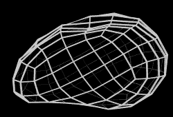
XCT-Only Sample
The XCT-Only models display only the unprocessed X-ray computed tomography image data of the interior of the rock. These models differ from our “fusion” models that display both the exterior high-resolution photogrammetric image data and the interior X-ray computed tomography data. Since the Astromaterials Research & Exploration Science CT Laboratory at NASA Johnson Space Center produces extensive image data for samples, Astromaterials 3D has created this special section to serve selections of this important data to researchers and the public. You will notice that the XCT image data shown here may have less contrast or look less in focus than in the “fusion” models—this is because the data here has not been adjusted at all. In the fusion model, our team adjusts the image data within certain acceptable scientific parameters to bring out details in the data, but what you see here is the unprocessed data as it is output by the XCT scanner. You are welcome to download this data for your own exploration and research—link below!
A note about the XCT-Only mesh visualization: The 3D mesh visible in this XCT-Only sample has very low fidelity to balance preservation of the general surface geometry while allowing a visually clear wireframe representation for our web application. Deviations between the XCT data and mesh geometry may occur as a result. The XCT data—not the mesh—is the definitive representation of the sample geometry.
A note about the XCT-Only mesh visualization: The 3D mesh visible in this XCT-Only sample has very low fidelity to balance preservation of the general surface geometry while allowing a visually clear wireframe representation for our web application. Deviations between the XCT data and mesh geometry may occur as a result. The XCT data—not the mesh—is the definitive representation of the sample geometry.
??? Processing Details
High-Resolution Precision Photography (HRPP)
Camera
Lens
Mega Pixels
Photos
Total Size
Photography
value
value
value
value
value
Structure-From-Motion Photogrammetry (SFM)
The SFM process is used to produce the 3D mesh of the rock’s exterior surface from the HRPP using photogrammetric principles. The rock’s texture is derived from HRPP and mapped to the mesh resulting in photorealistic color.
Micro X-ray Computed Tomography (XCT)
The XCT images allow you to look inside the rock itself, where the brightness variation of textural features you see is related to density and composition. For example, the bright tones would indicate metal or metal-rich features and the darker tones would indicate silica-rich features. Interior black features are voids.
Micro X-Ray Computed Tomography
Cut into the rock from three different orientations to reveal X-Ray CT imagery of the rock's interior.
Slice Orientation and Position
Use your mouse to drag the sliders below. Release the mouse for full resolution imagery.
 0cm
0cm
0cm
0cm
0cm
0cm
Details
Make fine selection changes with +- and open high resolution slice imagery.
View XCT Planes
XCT Slice Number
View Slice
XCT Slice Number
View Slice
-
0000
+
-
0000
+
-
0000
+
-
0000
+
-
0000
+
-
0000
+
View
Select a XCT scan orientation or nudge the view using the arrows.
Ambient
Light
Light
Spot
Light
Light
Anaglyph
3D
3D
Scale
Guide
Guide
Grid
Plane
Plane
Reset
View
View
Pins
The following pins were selected by NASA curators to showcase interesting features for each sample. Click the corresponding view button  to locate each pin.
Pin, comment on, and share points of interest within the sample. Click the corresponding view button to view each pin after creation.